
Regenerative Medicine Division
Info
-
Biofabrication Technologies
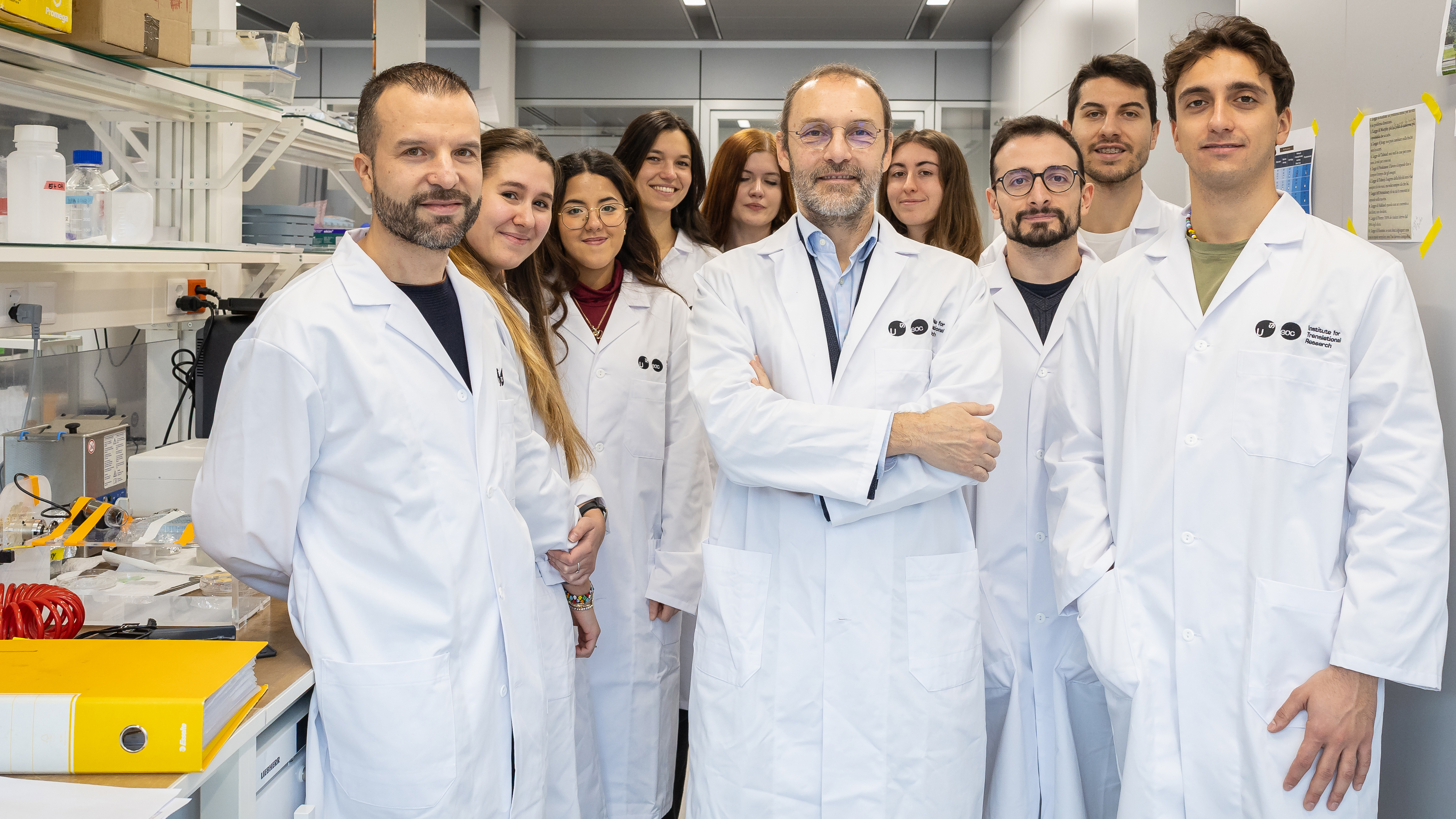
Prof Matteo Moretti, PhD
Principal Investigator
ORCID: 0000-0002-7301-1208Dalila Petta, Post doc
ORCID: 0000-0002-1412-0084Simone Gugliandolo, Post doc
ORCID: 0000-0002-9803-3761Giuseppe Talò
Engineer
ORCID: 0000-0003-1976-5073Alex Zadro, PhD student
ORCID: 0009-0003-1322-891XImen Dziri, PhD student
ORCID: 0009-0000-7629-5771Stella Monestier, PhD student
Andrea D’Angelo, visiting PhD student
Jacopo De Luca, Master student
Research area and translational research
The Regenerative Medicine Technologies (RMT) group adopts an interdisciplinary approach, at the intersection of engineering, biology and medicine, aiming at regenerating biological tissue substitutes in vitro. Major RMT competences are represented by advanced technologies such as biofabrication and microfluidics, enabling tools for the generation of microphysiological systems replicating the complexity of in vivo biology. In more detail, in RMT we generate 3D tissues based on cell co-cultures mainly of human origin, embedded in suitable biomaterials and cultured in engineered environments, such as bioreactors providing biophysical and mechanical stimuli. RMT mainly focuses on musculoskeletal tissues, producing biological replicates with a scale ranging from organ on chip devices to macro-scale constructs of clinically relevant dimensions. The microarchitecture of physiological tissues can be hierarchically replicated, spanning from mimicking the interface between different tissues (e.g. tissues composing the joints or the tendon bony insertion) down to the reproduction of microscale tissue features (e.g. bone trabeculae or perfusable microvascular networks).
From a translational point of view, microphysiological systems are currently being improved towards high throughput platforms usable for drug screening purposes and testing of personalized therapies, whilst clinically relevant constructs, including organoids, could be translated to biological substitutes usable in clinics for the regeneration of diseased tissues. Our more consolidated expertise is on musculoskeletal constructs, and recently we started addressing other tissue types including brain and neuromuscular tissues. Our aim is to leverage microphysiological systems and organoids to improve drug discovery and to personalize available therapies in fields such as musculoskeletal and degenerative diseases.
In RMT we developed patient-specific multi-tissue 3D models, such as an osteoarthritic joint on a chip, enabling the evaluation of different possible therapeutic approaches on the single patient for the development of personalized therapeutic regimens for osteoarthritis and other orthopedic pathologies. Our muscle models have shown organization of muscle fibers and presence of a vascular network similar to those in vivo, better reproducing the altered production of extracellular proteins in pathological conditions as compared to 2D models. In the field of bone tumors, we demonstrated that our models were able to reproduce the effects of known anti-tumor drugs on cancer cells better than simple 3D cancer cell models.
Finally, we recently started to leverage our competences in in vitro culture platform design to develop enabling technologies for biological studies in space. Microgravity and radiation, typical of the space environment, induce changes in biological systems like those induced by ageing at an accelerated pace, representing an interesting opportunity to study underlying mechanisms or to discover potential countermeasures. Since performing experiments in real microgravity requires to overcome logistic and engineering challenges, we setup a facility endorsed by European Space Agency (ESA) to help researchers interested in performing 3D cell cultures in space to organize their experimental setups.
Research methods
In the RMT Lab, innovative diagnostic and therapeutic methodologies are designed and developed, with particular reference to human 3D models in vitro that reproduce different tissues, musculoskeletal (muscle, bone and cartilage) and neuronal, vascularized when necessary.
The creation of miniaturized 3D models is based on the use of innovative technologies such as bio-manufacturing (including 3D printing and bio-printing) and microfluidics. A fundamental element of the 3D models is represented by cells, mostly of human origin, obtained commercially or directly isolated from surgical waste fragments.
To obtain the formation of 3D tissues, different cell populations of the reproduced tissue are co-cultured and incorporated into hydrogels injected into microfluidic or macroscale culture devices, which are custom developed in the laboratory.
The microphysiological systems developed can be used for the study of physio-pathological mechanisms or for the evaluation of drugs and therapies, by means of analyses mainly based on confocal and fluorescent imaging, as well as secretome analysis assays. Furthermore, we perform quantitative analyses on the cells extracted from the models themselves, such as transcriptomics and flow cytometry.
Finally, we have developed devices to simulate microgravity, allowing to perform experiments in conditions mimicking the space real microgravity.
Info
-
Vascular Aging
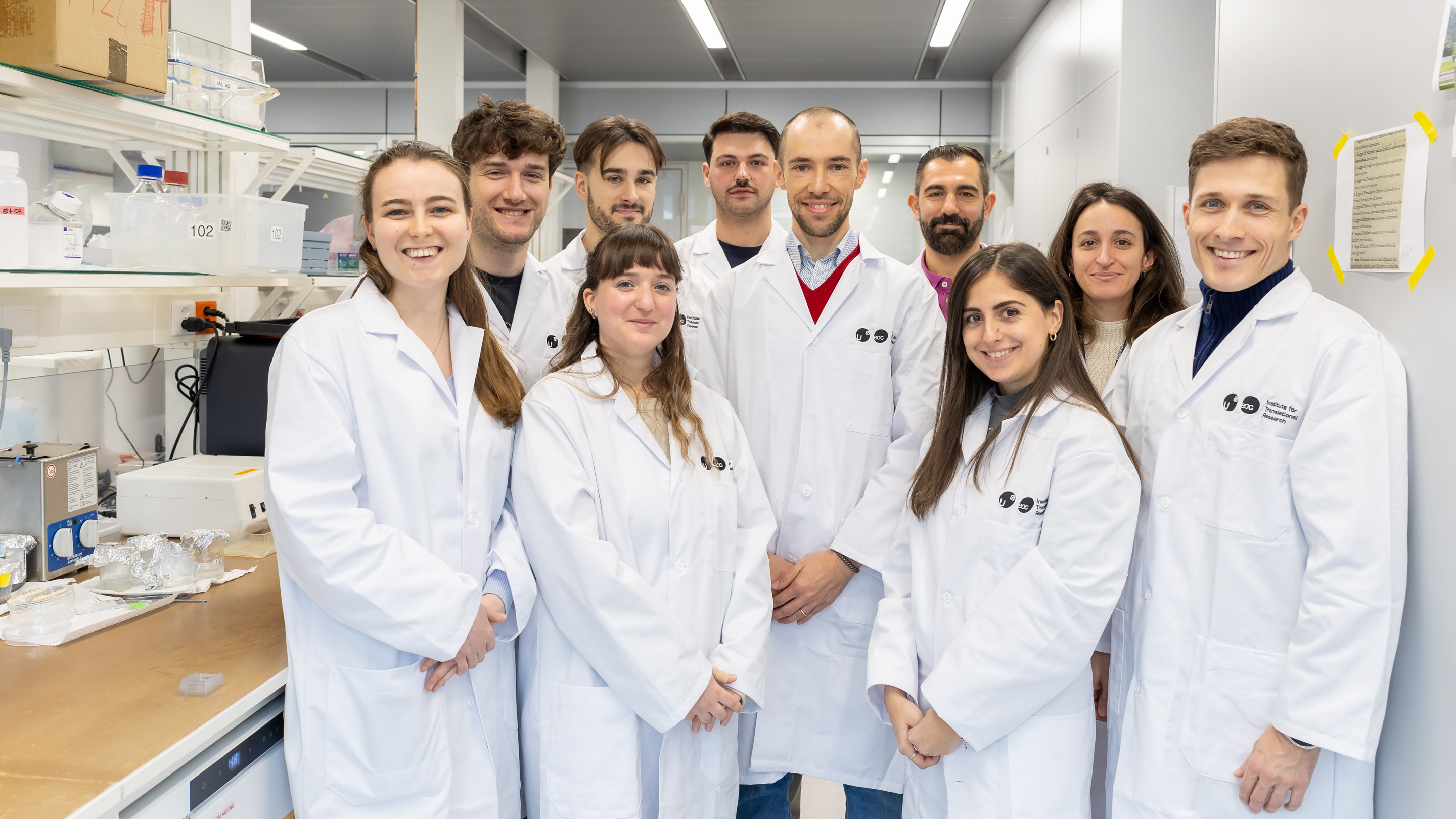
Prof Simone Bersini
Principal Investigator
ORCID: 0000-0003-4620-3500Andrea Uccelli, Post doc
ORCID: 0009-0001-2446-6740Francesco Gualdi, Post doc
ORCID: 0000-0003-0449-9884Filippo Zoppi, Post doc
Mattia Cenciarini, PhD student
ORCID: 0009-0000-0270-8537Chiara Zamboni, PhD student
ORCID: 0009-0004-6185-3151Dorian Tace, PhD student
ORCID: 0009-0008-1196-0396Julian Sackman, PhD student
Susanna Nodiroli, PhD student
Valentina Colombo, Research Assistant
Marika Natali, Master student
Research area and translational research
Identifying the biological mechanisms underlying blood vessel aging in order to prevent it, in an attempt to rejuvenate the surrounding tissues and organs: this is “vascular rejuvenation”, the goal of the five-year research project with which Simone Bersini won a 2.6 million Swiss franc ERC Starting Grant in 2023. The study combines microphysiological systems, omics analysis and machine learning, and could have implications not only in the cardiovascular field, but also in the fight against neurodegenerative diseases and tumour metastases. In the first phase of the project, miniaturised systems embedded within a high-throughput platform were developed and validated. These organ-on-a-chip allow to biofabricate in a few days perfusable capillary networks using patient-derived cells embedded in a 3D hydrogel matrix. A key aspect of this research line is the integration of automation, from tissue biofabrication to live imaging. This allows to reproducibly generate large number of samples that can be further analyzed through multi-omic techniques. Apart from molecular analyses, the biofabricated blood vessels are studied through functional assays (e.g. quantification of vascular leakage) to mechanistically connect gene/protein changes with a clear readout. A spin-off of the project is focused on the biofabrication of 3D lymphatic networks which are then exploited to compare how blood and lymphatic endothelia behave during aging.
This line of research has been further expanded in the following years to study the impact of sex-specific differences in vascular aging (Novartis FreeNovation) and to analyze the contribution of the aging blood brain barrier in cancer metastases (Swiss National Science Foundation, collaboration with Prof. Levesque at the University of Zurich). Merging an interdisciplinary background in bioengineering and molecular cell biology, the group is also focused on the design of novel strategies to promote organoid vascularization with the goal of understanding the contribution of blood vessels in brain development and disease (Swiss National Science Foundation, collaboration with Prof. Baggiolini at the Institute of Oncology Research). Last but not least, the group is devoted to the study of vascular dysfunctions in the onset and progression of neuro-muscular diseases, focusing on the contribution of the endothelium. Custom organ-on-a-chip are being developed to couple contractile human muscle fibers with capillary networks with the goal of clarifying the complex cellular interactions occurring within pathological muscle microenvironments (Fondation suisse de recherche sur les maladies musculaires and Novartis Foundation for Medical-Biological Research).
Research methods
The group combines the design of high-throughput culture systems with the biofabrication of 3D miniaturized models of perfusable blood vessels, which are then integrated into organ-specific microenvironments (e.g. brain, skeletal muscle, bone) according to the specific biological question. These organs-on-a-chip include cells of human origin which can be isolated from patient biopsies to create personalized models. The systems are exposed to physiological stimulation (e.g. luminal and interstitial flow) and can be monitored through molecular (e.g. RNA sequencing, mass spectrometry, multiplex secretome) and functional analyses (e.g. blood vessel permeability, muscle contractility). Artificial intelligence-based methods are developed in collaboration with IDSIA to move beyond correlative analyses and identify key determinants of specific patho-physiological processes.
Info
-
Tumor microenvironment
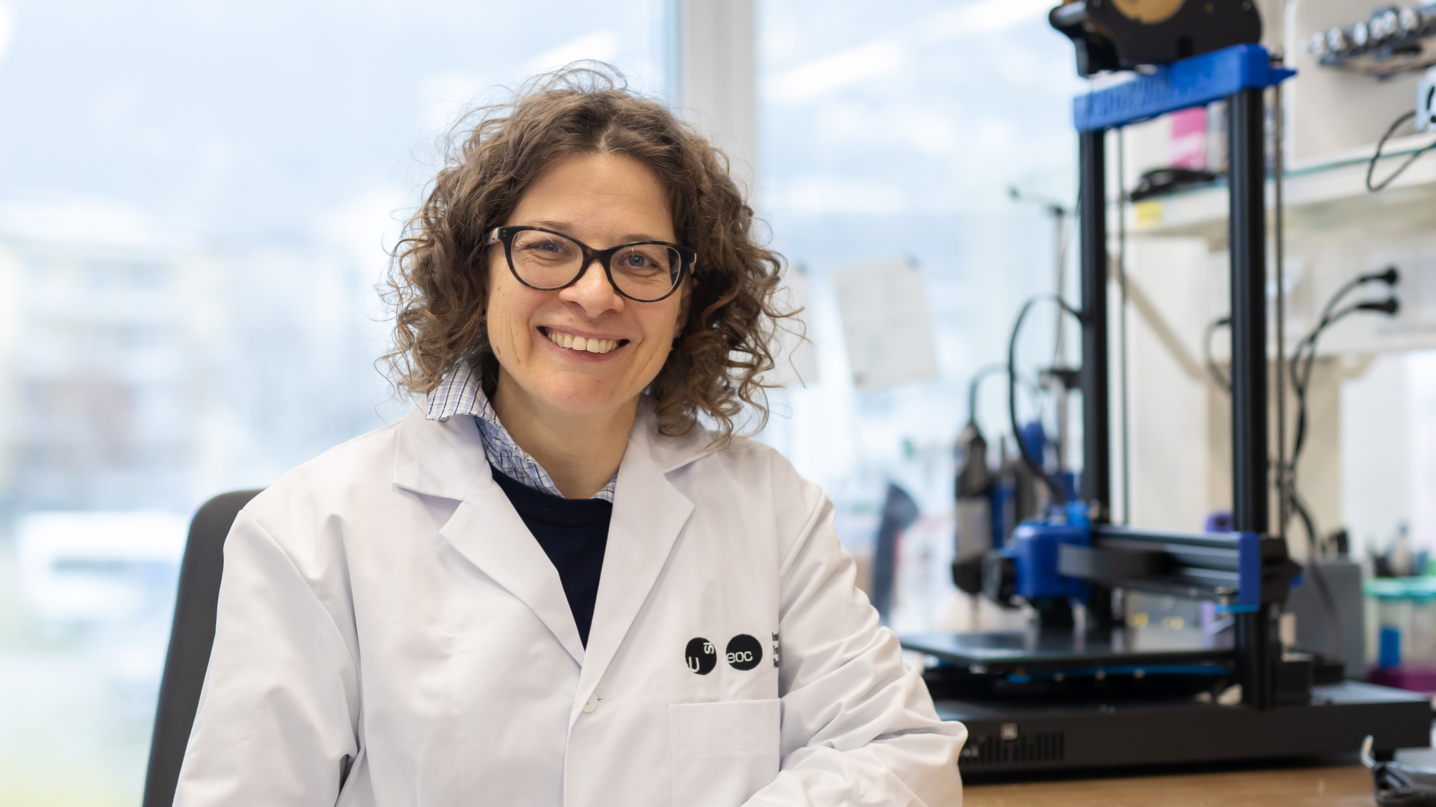
PD Chiara Arrigoni
Principal Investigator
ORCID: 0000-0001-6808-1642
Ewelina Latoszek, Post doc
ORCID: 0000-0002-7883-6989
Viviana Secci, PhD student
ORCID: 0009-0002-7370-9419Research area and translational research
One of the most dangerous features of malignant tumors is their propensity to spread from the original site towards secondary organs, forming metastases which are presently incurable, representing the major cause of death. Several factors are involved in the metastatization process, and microenvironmental niches in the secondary organs have been indicated as one of the crucial aspects determining the establishment of metastases in specific tissues. In particular, bone and bone marrow are frequent sites of metastatic growth for different types of tumors, suggesting that intrinsic features of the bone environment can be critical determinants for secondary tumor establishment. In this context, there is a pressing need for a better understanding of the metastatic cascade and for the discovery of new effective tehrapies counteracting this phenomenon. The discovery of new therapeutic approaches is a challenging process, in which several candidate molecules, identified in the preclinical phase, fail due to safety or efficacy issues. This is mainly due to the inadequacy of presently available preclinical models, represented by standard 2D cultures or animal models. The aim of our research is thus to develop more reliable in vitro models replicating the architecture and composition of the metastatic tumor environment, based on human cells and leveraging biofabrication and organ-on-chip technologies. These advanced models will be exploited for evaluating new drug efficacy and safety in a more reliable way and will enable the implementation of patient-tailored assays, in line with a personalized medicine approach.
Ongoing projects are focused on the bone environment and its role in tumors such as breast cancer, prostate cancers, sarcoma and lymphomas. For the study of sarcoma progression, we developed a complex multicellular model replicating the process of endochondral ossification and showing how different developmental stages differently influenced tumor cell growth. We are now developing bone on chip models with mechanical properties of the support matrix similar to native bone, exploiting bioprinting techniques. In parallel, we are generating bone marrow-like microphysiological systems with high throughput to enable the screening of drugs against lymphoma and prostate cancer, to help identifying the most effective therapies.
Research methods
To generate reliable models of the tumor microenvironment, we co-culture multiple types of human cells, mainly primary cells derived from human tissues harvested from patients upon informed consent. Three-dimensional architecture of the tissue is provided by the use of extracellular matrices, embedding cell cultures. To more faithfully replicate the geometry of the native tissue, we leverage technologies including microfabrication and 3D printing.
Furthermore, to reproduce tissue-specific biophysical cues, such as interstitial perfusion or mechanical stimulation, microfluidic and organ on chip technologies are exploited.
From an analytical perspective, 3D in vitro models are particularly suited for spatial analyses including fluorescence and confocal imaging. Assays including analyses of the secretome and transcriptomics are also applied to culture medium or to cells extracted from the models.
Info
-
Recent Publications
- A high-throughput bone marrow 3D co-culture system to study resistance to BCR signalling targeted agents in B-NHL. Zadro A, Arribas A, Colombo MV, Cannas E, Spriano F, Cascione L, Mensah AA, Simonetta F, Petta D, Candrian C, Arrigoni C, Bertoni F, Moretti M. Br J Haematol. 2025 Dec 8. doi: 10.1111/bjh.70273. Online ahead of print.PMID: 41362022
- Biocompatible Ink Optimization Enables Functional Volumetric Bioprinting With Xolography. Brauer E, Balciunaite A, Kollert MR, Weihs J, Knecht RS, Behncke R, Quach S, Felix König N, Badolato A, Monestier S, Bersini S, Moretti M, Filippi M, Hägerling R, Rezvani M, Hecht S, Petersen A, Katzschmann RK.Adv Mater. 2025 Nov 29:e12058. doi: 10.1002/adma.202512058. Online ahead of print.PMID: 41318943
- A Compartmentalized Joint-on-chip (JoC) Model to Unravel the Contribution of Cartilage and Synovium to Osteoarthritis Pathogenesis. Palma C, Salehi S, Polidoro MA, Moretti M, Rasponi M, Lopa S, Occhetta P.Adv Sci (Weinh). 2025 Nov;12(42):e00374. doi: 10.1002/advs.202500374. Epub 2025 Sep 11.PMID: 40936111
- Impact of adipose-derived mesenchymal stem cells and their secretome on osteoarthritis in a rat model. Palombella S, Lopa S, Recordati C, Canesi S, Moretti M, Lovati AB.BMC Musculoskelet Disord. 2025 Apr 21;26(1):392. doi: 10.1186/s12891-025-08642-8.PMID: 40259333
- Matrix-assisted autologous chondrocyte transplantation is effective at mid/long-term for knee lesions: A systematic review and meta-analysis. Colombini A, Raffo V, Gianola S, Castellini G, Filardo G, Lopa S, Moretti M, de Girolamo L.Knee Surg Sports Traumatol Arthrosc. 2025 Aug;33(8):2866-2879. doi: 10.1002/ksa.12549. Epub 2024 Dec 3.PMID: 39624924
- Microvascular Health as a Key Determinant of Organismal Aging. Cenciarini M, Uccelli A, Mangili F, Grunewald M, Bersini S.Adv Sci (Weinh). 2025 Nov 5:e08659. doi: 10.1002/advs.202508659. Online ahead of print.PMID: 41194423
- Biofabrication of a 3D human skeletal muscle microenvironment to study the early steps of fibrosis. Francescato R, Ishmaku M, Talò G, Francese M, Cascione L, Martini V, Uguccioni M, Moretti M, Bersini S.Mater Today Bio. 2025 Oct 15;35:102386. doi: 10.1016/j.mtbio.2025.102386. eCollection 2025 Dec.PMID: 41158711
- Development of a Microfluidic Vascularized Osteochondral Model as a Drug Testing Platform for Osteoarthritis. Salehi S, Brambilla S, Rasponi M, Lopa S, Moretti M. Adv Healthc Mater. 2024 Oct 6:e2402350. doi: 10.1002/adhm.20240235
- A personalized osteoarthritic joint-on-a-chip as a screening platform for biological treatments. Petta D, D’Arrigo D, Salehi S, Talò G, Bonetti L, Vanoni M, Deabate L, De Nardo L, Dubini G, Candrian C, Moretti M, Lopa S, Arrigoni C. Materials Today Bio, 2024, 26,101072
- Complex or not too complex? One size does not fit all in next generation microphysiological systems. Bersini S, Arrigoni C, Talò G, Candrian C, Moretti M. iScience. 2024 Feb 12;27(3):109199. doi: 10.1016/j.isci.2024.109199
- Endothelial-mesenchymal transition in skeletal muscle: Opportunities and challenges from 3D microphysiological systems. Francescato R, Moretti M, Bersini S. Bioeng Transl Med 2024 doi.org/10.1002/btm2.10644
- Differential angiogenesis of bone and muscle endothelium in aging and inflammatory processes. Arrigoni, C., Ostano, P., Bersini, S. Crippa M, Colombo MV, Gilardi M, Zagra L, Mello-Grand M, Gregnanin I, Ghilardi C, Bani MR, Candrian C, Chiorino G, Moretti M. Commun Biol 6, 126 (2023).
- In vitro models of breast cancer bone metastasis: analyzing drug resistance through the lens of the microenvironment Lamouline A, Bersini S, Moretti M., Frontiers in Oncology 13, (2023)
- A microfluidic model of human vascularized breast cancer metastasis to bone for the study of neutrophil-cancer cell interactions. Crippa M, Talò G, Lamouline A, Bolis S, Arrigoni C, Bersini S, Moretti M. Mater Today Bio. 2022 Oct 10;17:100460
- Culture of 3D Bioprinted Bone Constructs Requires an Increased Fluid Dynamic Stimulation Mainardi V.L., Sabato C., Rubert M., de Leeuw A., Arrigoni C., Dubini G., Candrian C., Moretti M, Müller R., Acta Biomaterialia 2022 Sep 13;S1742-7061(22)00573-6.
- Assessing the response of human primary macrophages to defined fibrous architectures fabricated by melt electrowriting Mondadori C, Chandrakarb A, Lopa S, Wieringa P, Talò G, Perego S, Lombardi G, Colombini A, Moretti M. Bioactive Materials 2022 Aug 27;21:209-222
- Musculoskeletal tissues-on-a-chip: role of natural polymers in reproducing tissue-specific microenvironments Petta D, D’Amora U, D’Arrigo D, Tomasini M, Moretti M. Biofabrication 2022 Aug 31;14(4).
- Integrative gene network and functional analyses identify a prognostically relevant key regulator of metastasis in Ewing sarcoma Cidre-Aranaz F, Li J, Hölting TLB, Orth MF, Imle R, Kutschmann S, Ammirati G, Ceranski K, Carreño-Gonzalez MJ, Kasan M, Marchetto A, Funk CM, Bestvater F, Bersini S, Arrigoni C, Moretti M, Thiel U, Baumhoer D, Sahm F, Pfister SM, Hartmann W, Dirksen U, Romero-Pérez L, Banito A, Ohmura S, Musa J, Kirchner T, Knott MML, Grünewald TGP.. Mol Cancer. 2022 Jan 3;21(1):1
- Recapitulating monocyte extravasation to the synovium in an organotypic microfluidic model of the articular joint. Mondadori C, Palombella S, Salehi S, Talò G, Visone R, Rasponi M, Redaelli A, Sansone V, Moretti M, Lopa S. Biofabrication. 2021 Jul 7;13(4).
- The driving role of the Cdk5/Tln1/FAKS732 axis in cancer cell extravasation dissected by human vascularized microfluidic models Gilardi M, Bersini S, Valtorta S, Proietto M, Crippa M, Boussommier-Calleja A, Labelle M, Moresco RM, Vanoni M, Kamm RD, Moretti M.. Biomaterials. 2021 Jul 20;276:120975
- Engineering the early bone metastatic niche through human vascularized immuno bone minitissues. Colombo MV, Bersini S, Arrigoni C, Gilardi M, Sansoni V, Ragni E, Candiani G, Lombardi G, Moretti M. Biofabrication. 2021 Apr 26;13(3).
- Radiobiological Studies of Microvascular Damage through In Vitro Models: A Methodological Perspective. Possenti L, Mecchi L, Rossoni A, Sangalli V, Bersini S, Cicchetti A, Costantino ML, Candrian C, Arrigoni C, Rancati T, Moretti M. Cancers (Basel). 202
- Improving cell seeding efficiency through modification of fiber geometry in 3D printed scaffolds. Mainardi VL, Arrigoni C, Bianchi E, Talò G, Delcogliano M, Candrian C, Dubini G, Levi M, Moretti M. Biofabrication. 2021
- A microphysiological early metastatic niche on a chip reveals how heterotypic cell interactions and inhibition of integrin subunit β3 impact breast cancer cell extravasation Crippa M, Bersini S, Gilardi M, Arrigoni C, Gamba S, Falanga A, Candrian C, Dubini G, Vanoni M, Moretti M.. Lab Chip. 2021 Mar 21;21(6):1061-1072
- Sound-induced morphogenesis of multicellular systems for rapid orchestration of vascular networks. Petta D, Basoli V, Pellicciotta D, Tognato R, Barcik JP, Arrigoni C, Della Bella E, Armiento AR, Candrian C, Richards GR, Alini M, Moretti M, Eglin D, Serra T. Biofabrication. 2020 Sep 25
- Organs-on-a-chip as model systems for multifactorial musculoskeletal diseases. Arrigoni C, Lopa S, Candrian C, Moretti M. Curr Opin Biotechnol. 2020 Jun;63:79-88.
- Oncogenic hijacking of a developmental transcription factor evokes vulnerability toward oxidative stress in Ewing sarcoma Marchetto A, Ohmura S, Orth MF, Knott MML, Colombo MV, Arrigoni C, Bardinet V, Saucier D, Wehweck FS, Li J, Stein S, Gerke JS, Baldauf MC, Musa J, Dallmayer M, Romero-Pérez L, Hölting TLB, Amatruda JF, Cossarizza A, Henssen AG, Kirchner T, Moretti M, Cidre-Aranaz F, Sannino G, Grünewald TGP.. Nat Commun. 2020 May 15;11(1):2423.
- Microvascular Health as a Key Determinant of Organismal Aging. Cenciarini M, Uccelli A, Mangili F, Grunewald M, Bersini S.Adv Sci (Weinh). 2025 Dec;12(47):e08659. doi: 10.1002/advs.202508659. Epub 2025 Nov 5.PMID: 41194423
- Hetzer MW and Bersini S, Beyond Static Pipes: Mechanisms and In Vitro Models of Vascular Aging, Cold Spring Harb Perspect Med 2022, a041180. doi: 10.1101/cshperspect.a041180 https://perspectivesinmedicine.c7shlp.org/content/12/6/a041180
- Bersini S, Schulte R, Huang L, Tsai H, Hetzer MW. Direct reprogramming of human smooth muscle and vascular endothelial cells reveals defects associated with aging and Hutchinson-Gilford Progeria Syndrome. eLife 2020, 9:e54383 https://elifesciences.org/articles/54383
- Bersini S, Arrojo e Drigo R, Huang L, Shokhirev MN, Hetzer MW. Transcriptional and functional changes of the human microvasculature during physiological aging and Alzheimer Disease. Advanced Biosystems 2020, 4(5):e2000044. doi: 10.1002/adbi.202000044 https://onlinelibrary.wiley.com/doi/full/10.1002/adbi.202000044
- Bersini S, Lytle NK, Schulte R, Huang L, Wahl GM, Hetzer MW. Nup93 regulates breast tumor growth by modulating cell proliferation and actin cytoskeleton remodeling. Life Science Alliance 2020, 3(1) pii:e201900623. doi: 10.26508/lsa.201900623 https://www.life-science-alliance.org/content/3/1/e201900623
